
















Advanced Microscope Could Bring New Hope for IBD Patients
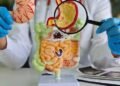
A cutting-edge microscope could help scientists better understand the biological processes behind inflammatory bowel disease (IBD), raising the possibility of developing more precise treatments for people living with the long-term condition.
Researchers at the Rosalind Franklin Institute said the new super-resolution imaging technology can produce images at roughly 10 times the resolution of conventional light microscopes. Scientists hope the technique will provide new insights into how IBD develops and help identify potential approaches to treatment.
Technology Designed to Reveal Hidden Changes
The new Stimulated Emission Depletion (STED) microscope is based at the Harwell Science and Innovation Campus in Oxfordshire. It uses a second laser with a doughnut-shaped beam alongside the main laser.
The outer section of the second laser suppresses fluorescence generated around the main beam, while its centre remains unaffected. This enables researchers to capture fluorescent signals from the precise focal point with much greater detail.
STED microscopy can also examine several types of molecules simultaneously. This multiplexing capability allows researchers to label biomolecules, including proteins, lipids and glycans, with fluorescent markers so that multiple structures can be distinguished within the same sample.
The UK’s only microscope of this type also contains features designed to improve imaging and allow more realistic biological studies. Special mirrors compensate for image distortion, while a temperature-controlled platform makes it possible to examine living cells and mini-organs at temperatures close to those inside the human body.

The combination of these capabilities means scientists can observe living samples in real time or examine preserved samples in detail. Researchers believe this could help uncover the cellular and molecular processes associated with IBD and the symptoms it can cause.
The project is being led by Dr Karina Pombo-Garcia, whose team hopes the technology will eventually contribute to better care for the more than 500,000 people in the UK living with IBD. Crohn’s disease and ulcerative colitis are the condition’s two primary forms.
Dr Pombo-Garcia told EMJ that STED provides researchers with the ability to examine the molecular structure of the human gut at about 20 nanometres, offering approximately 10 times greater resolution than conventional light microscopy.
At this level of detail, researchers can investigate how individual proteins are positioned at the connections between intestinal cells and how they contribute to the gut’s protective barrier.
This could be particularly important for IBD research because the intestinal barrier becomes damaged and more permeable in people with the disease, which can allow inflammation to continue. Scientists, however, do not yet have a complete picture of the microscopic structural changes responsible for this damage.
Traditional microscopy can demonstrate that the intestinal barrier has been disrupted, but the researchers believe STED could provide a much more precise picture of where the disruption occurs and what causes it.
The technology can also be applied to thicker human tissue samples, including intestinal organoids created from patients. By studying healthy organoids alongside those derived from people with IBD, the team hopes to identify the earliest molecular changes that contribute to failure of the gut barrier.
Funding Boosts UK Research Efforts
The development comes amid continuing concerns about the impact of IBD on patients. Research cited by the British Society of Gastroenterology found that almost a quarter, or 24%, of UK adults living with IBD have difficulty coping with the disease. The report also indicated that approximately one in seven cases are diagnosed after an emergency hospital admission.
For many patients, effective targeted treatment options remain limited, while some existing therapies can produce debilitating side effects that interfere with daily life.
Researchers believe the new imaging technology could help address this gap by improving understanding of the molecular changes involved in IBD.
Dr Pombo-Garcia said identifying the precise molecular processes that go wrong is essential for developing genuinely targeted therapies. She explained that STED could help researchers discover possible treatment targets and, eventually, determine whether therapies can restore the gut barrier to its normal nanoscale organization.
Rather than simply providing higher-quality images, she said, the technology offers a new method for measuring aspects of human disease biology that previously could not be seen.
Science Minister Chris McDonald officially unveiled the microscope alongside a five-year investment of £67.7 million from the Engineering and Physical Sciences Research Council (EPSRC) for the Rosalind Franklin Institute.
The funding will support the institute’s research into advanced imaging, engineering, biology and diagnostic technologies.
Dr Pombo-Garcia said the new funding would give her team an exceptional opportunity to advance its research and technology development. She said it could allow IBD researchers to link changes occurring at the level of individual proteins with the effects ultimately experienced by patients.
The Henry Royce Institute, which specializes in materials research, is also due to receive an additional £90 million over the same period. The investment forms part of the government’s broader effort to strengthen scientific and medical innovation in the UK.
The funding was included in the core UK Research and Innovation (UKRI) budget announced as part of the 2025 spending review.
For more health-related news